Eye Diseases: Diagnosis, Treatment, and Safety Guide
A clear guide to common eye diseases, warning signs, early diagnosis, and treatment options for cataract, glaucoma, AMD, and diabetic retinopathy.
Introduction
Some of the most serious eye diseases do not begin with pain or dramatic symptoms. They may progress quietly, and by the time blurred vision, reading difficulty, or loss of visual field appears, part of the damage may already be difficult to reverse.
The central message of this article is simple: timely eye examinations can protect vision. This is especially important for older adults, people with diabetes, high blood pressure, a family history of glaucoma, high myopia, long-term steroid use, or new visual symptoms.
Instead of treating eye disease as a long list of diagnoses, it is useful to group conditions into three practical categories:
| Category | Examples | What to remember |
|---|---|---|
| Common and usually gradual | Cataract, dry eye, changes in glasses prescription | Often not an emergency, but follow-up is needed when function is affected |
| Potentially silent and progressive | Glaucoma, diabetic retinopathy, early AMD | Periodic examination matters even without pain or major symptoms |
| Urgent conditions | Retinal detachment, retinal vascular occlusion, severe infection, painful red eye with vision loss | Same-day evaluation or emergency eye care may be required |
This article presents the major eye diseases in a structured way: what the problem is, how it may feel, how it is diagnosed, what treatments exist, and when urgent care is needed.
Causes and Symptoms
Below is a structured overview of common eye diseases by mechanism, typical symptoms, and main risk.
Cataract
What happens? The natural lens becomes cloudy, most often as part of aging. Common symptoms: Gradual blurred vision, glare at night, difficulty driving, and frequent changes in glasses prescription. Risk factors: Age, diabetes, long-term sun exposure, smoking, prolonged steroid use, and eye trauma. Key point: Cataract is usually not an emergency, but when it interferes with daily function, surgery is often highly effective.
Glaucoma
What happens? Progressive damage to the optic nerve, often associated with elevated intraocular pressure. Common symptoms: Early disease usually has no symptoms. Later, peripheral visual field loss may occur. Risk factors: Age, family history, elevated intraocular pressure, high myopia, certain ancestry backgrounds, and steroid use. Key point: Glaucoma is sometimes called the “silent thief of sight” because damage may progress without pain or obvious central vision loss.
Age-Related Macular Degeneration (AMD)
What happens? The central retina, responsible for reading, face recognition, and fine detail, is affected. Common symptoms: Distorted straight lines, reading difficulty, a central spot, or reduced central vision. Risk factors: Age, smoking, family history, sun exposure, and a diet low in protective nutrients. Key point: In wet AMD, early anti-VEGF injections may help preserve vision and prevent major deterioration.
Diabetic Retinopathy
What happens? Diabetes damages small retinal blood vessels and may cause bleeding, swelling, and vision loss. Common symptoms: Early stages may cause no symptoms. Later, blurred vision, floaters, or reduced vision may occur. Risk factors: Duration of diabetes, poor glucose control, high blood pressure, kidney disease, and pregnancy in women with diabetes. Key point: Periodic retinal examination is important even when vision feels normal.
Keratoconus
What happens? The cornea becomes thinner and more cone-shaped, causing irregular focusing of light. Common symptoms: Rapid prescription changes, distorted or double vision, night-driving difficulty, and glare. Risk factors: Family history, eye rubbing, allergic eye disease, and onset at a young age. Key point: Early diagnosis allows treatments such as corneal cross-linking, which may slow or stop progression.
Dry Eye and Eyelid Inflammation
What happens? Tear quantity or tear quality is reduced, often with inflammation of the eyelid margins. Common symptoms: Burning, gritty sensation, redness, reflex tearing, screen-related fatigue, and foreign-body sensation. Risk factors: Prolonged screen use, age, certain medications, autoimmune disease, and dry environments. Key point: Most cases are uncomfortable rather than dangerous, but redness with pain and vision loss requires examination.
Types of Eye Disease Compared
| Disease | Main structure affected | Typical sign | Usually painful? | Urgency level |
|---|---|---|---|---|
| Cataract | Lens | Gradual blur and glare | Usually no | Follow-up or surgery depending on function |
| Glaucoma | Optic nerve | Peripheral visual field loss | Usually no | Periodic testing; urgent if acute pain and redness occur |
| AMD | Central retina | Distorted lines and central vision loss | Usually no | Urgent if sudden change occurs |
| Diabetic retinopathy | Retinal blood vessels | Often silent until advanced | Usually no | Regular screening; urgent if sudden vision loss occurs |
| Keratoconus | Cornea | Rapid prescription changes and distorted vision | Usually no | Prompt assessment, especially in younger patients |
| Dry eye | Tear film / ocular surface | Burning, gritty sensation, screen fatigue | Sometimes burning | Usually not urgent |
| Retinal detachment | Retina | Flashes, floaters, dark curtain | Usually no | Eye emergency |
| Corneal infection | Cornea | Pain, redness, light sensitivity, vision loss | Yes | Very urgent |
This comparison helps separate common discomfort from potentially sight-threatening symptoms. Practical rule: sudden vision loss, strong pain, flashes of light, or a curtain-like shadow in the visual field should not be ignored.
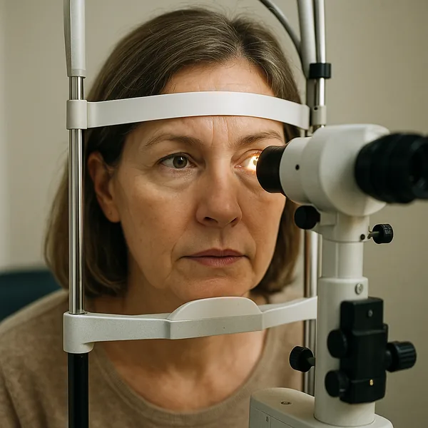
Diagnosis
Diagnosis of eye disease combines the patient’s symptoms, clinical examination, and imaging. Not every test is needed for every person; the choice depends on age, symptoms, and risk factors.
| Test | What it checks | When it is especially useful |
|---|---|---|
| Visual acuity test | Ability to see detail near and far | Blurred vision, prescription changes, general assessment |
| Slit-lamp examination | Cornea, lens, eyelids, and front of the eye | Cataract, dry eye, inflammation, foreign body |
| Intraocular pressure measurement | Pressure inside the eye | Glaucoma suspicion or monitoring |
| Dilated fundus examination | Retina, blood vessels, and optic nerve | Diabetes, AMD, flashes, reduced vision |
| OCT | Detailed cross-sectional imaging of retina and optic nerve | AMD, macular edema, glaucoma |
| Visual field test | Peripheral visual function | Glaucoma and optic nerve disease |
| Corneal topography | Shape and curvature of the cornea | Keratoconus, contact lens fitting, laser surgery evaluation |
Important principle
One normal examination does not protect someone for life. Some eye diseases develop gradually, so repeat follow-up according to the eye doctor’s recommendation is often more valuable than a single test.
Treatment
Treatment depends on the diagnosis, severity, age, and impact on daily function. There is no single treatment that fits all eye diseases.
| Disease | Common treatment | Treatment goal |
|---|---|---|
| Cataract | Removal of the cloudy lens and implantation of an intraocular lens | Improve visual clarity and reduce glare |
| Glaucoma | Pressure-lowering drops, laser, or surgery when needed | Slow optic nerve damage |
| Dry AMD | Monitoring, smoking cessation, appropriate nutrition, and sometimes AREDS supplements when recommended | Slow progression |
| Wet AMD | Intravitreal anti-VEGF injections | Reduce abnormal vessel leakage and preserve vision |
| Diabetic retinopathy | Glucose and blood pressure control, laser, anti-VEGF, or surgery in advanced cases | Prevent bleeding, swelling, and vision loss |
| Keratoconus | Glasses, specialty contact lenses, cross-linking, and sometimes corneal transplant | Improve vision and slow progression |
| Dry eye | Artificial tears, eyelid hygiene, inflammation treatment, and sometimes prescription medications | Improve comfort and protect the ocular surface |
What not to do without medical advice
Do not use steroid drops, antibiotic drops, or old eye drops left at home without an examination. The wrong treatment can worsen infection, raise intraocular pressure, or hide important warning signs.
Multisystem Impact
The eyes are not isolated from the rest of the body. General diseases, medications, and lifestyle factors can directly affect the retina, optic nerve, cornea, and ocular blood vessels.
Diabetes
Diabetes can damage small retinal blood vessels. A person may still see well while early retinal damage is present, so periodic retinal screening is important.
High Blood Pressure and Vascular Disease
Uncontrolled blood pressure can appear as changes in retinal blood vessels and may increase the risk of vascular occlusion in the eye.
Autoimmune Disease
Inflammatory diseases may cause severe dry eye, intraocular inflammation, or optic nerve involvement.
Medications
Some medications can affect the eye. For example, steroids may raise intraocular pressure or accelerate cataract development. Patients should tell their eye doctor about regular medications.
Smoking
Smoking is a major risk factor for AMD and vascular disease progression. Stopping smoking is also an eye-health intervention.
Warning Signs
The following signs are not meant to frighten readers; they help distinguish ordinary discomfort from potentially sight-threatening symptoms.
| Warning sign | Possible meaning | What to do |
|---|---|---|
| Sudden vision loss in one or both eyes | Vascular occlusion, bleeding, optic nerve inflammation, retinal problem | Seek same-day urgent care |
| New flashes of light or many new floaters | Retinal tear or detachment | Urgent eye examination |
| Curtain-like shadow or dark area in the visual field | Possible retinal detachment | Emergency eye care |
| Strong eye pain with redness and vision loss | Infection, acute glaucoma, or severe inflammation | Urgent examination |
| Eye pain with nausea, halos around lights, or severe headache | Possible acute rise in intraocular pressure | Emergency eye care |
| Severe light sensitivity with contact lens use | Possible corneal infection | Remove lenses and seek urgent care |
| Sudden distortion of straight lines | Wet AMD or macular disease | Prompt examination |
Safety rule: significant pain + redness + reduced vision should not be managed with home drops or delayed for several days.
When to See a Doctor
Same-day urgent evaluation
Seek urgent care for sudden vision loss, new flashes of light, a curtain-like shadow, strong eye pain, major redness with reduced vision, eye injury, or unusual symptoms in contact lens users.
Prompt appointment, but not necessarily emergency care
Book a near-term examination for persistent blur, rapid prescription changes, worsening glare, reading difficulty, distorted lines, or dry-eye symptoms that do not improve.
Periodic follow-up
| Group | General follow-up to consider |
|---|---|
| Adults without specific complaints | Based on age and clinician recommendation |
| People with diabetes | Periodic retinal examination according to medical guidance |
| Family history of glaucoma | Intraocular pressure, optic nerve, and visual field testing as advised |
| Older age or suspected AMD | Retinal monitoring and sometimes OCT |
| High myopia | Retinal examination, especially with flashes or new floaters |
Practical principle: a new and rapid symptom needs a rapid response; a stable ongoing issue needs organized follow-up.
Practical Tips
Daily eye protection
- Wear sunglasses with reliable UV protection during sun exposure.
- Do not smoke, especially when AMD risk is present.
- Avoid forceful eye rubbing, particularly in young people or those at risk for keratoconus.
- Keep glucose and blood pressure controlled when diabetes or hypertension is present.
- Remove contact lenses if pain, redness, or reduced vision occurs.
Screen work
- Screens usually do not “destroy” the eyes, but they can worsen dryness and fatigue.
- Blink more often, take short breaks, and adjust lighting.
- Persistent blur, headaches, or reading difficulty should prompt evaluation for prescription changes and dry eye.
Medications and drops
- Do not use steroid drops without medical supervision.
- Redness-relief drops are not a long-term solution and may hide a problem.
- Artificial tears can help dry eye but do not replace an examination when pain or vision loss is present.
FAQ
Does screen work permanently damage vision?
Usually no. Prolonged screen use can cause dryness, fatigue, burning, and temporary blur, but it usually does not cause permanent eye damage. If blur persists after rest, an examination is recommended.
What is the difference between cataract and glaucoma?
Cataract is clouding of the lens and is often treated with surgery that improves vision. Glaucoma is damage to the optic nerve, and treatment mainly aims to stop or slow further damage; lost vision is not always recoverable.
Is glaucoma always painful?
No. Most chronic glaucoma progresses without pain. Severe pain with redness and vision loss may indicate an acute emergency, but chronic glaucoma can progress silently.
Can diabetes damage the eyes even when vision feels normal?
Yes. Diabetic retinopathy can begin without symptoms. Periodic retinal examinations are important even when vision seems normal.
When is distortion of straight lines concerning?
If straight lines look wavy, broken, or distorted, especially in one eye, a prompt eye examination is recommended. This may indicate macular disease, including wet AMD.
Is dry eye dangerous?
Most dry eye is uncomfortable rather than dangerous. However, severe pain, significant redness, light sensitivity, or reduced vision are not typical dry-eye symptoms and require examination.
Can all eye diseases be prevented?
No. Some conditions are related to age, genetics, or underlying disease. However, periodic examinations, diabetes and blood-pressure control, smoking cessation, and UV protection can reduce risk or allow earlier treatment.
Summary
Eye disease is a broad field, but it can be understood through one central message: not every dangerous eye disease feels dangerous at first. Cataract may develop gradually, glaucoma may damage peripheral vision without pain, diabetic retinopathy may progress while vision still seems normal, and AMD may begin with subtle changes in central vision.
This article is organized to help readers distinguish common discomfort from warning signs that require urgent care. Sudden vision loss, strong pain, flashes of light, a curtain-like shadow, or redness with reduced vision should be evaluated quickly.
The most important insight is that eye examinations are not only for people who already see poorly. For people with diabetes, older age, family history of glaucoma, or new visual symptoms, timely examination can be the difference between manageable treatment and irreversible damage.
References
- World Health Organization. Vision Impairment and Blindness Fact Sheet. 2024. https://www.who.int/news-room/fact-sheets/detail/blindness-and-visual-impairment
- American Academy of Ophthalmology. Age-Related Macular Degeneration Preferred Practice Pattern. 2024. https://www.aao.org/education/preferred-practice-pattern/age-related-macular-degeneration-ppp
- Gazzard G, et al. Selective laser trabeculoplasty versus eye drops for first-line treatment (LiGHT). Lancet. 2019;393:1505-16. https://www.thelancet.com/journals/lancet/article/PIIS0140-6736(18)32213-X/fulltext
- Heier JS, et al. Faricimab for neovascular AMD (TENAYA and LUCERNE). Lancet. 2022;399:729-40. https://www.thelancet.com/journals/lancet/article/PIIS0140-6736(22)00010-1/fulltext
- Chew EY, et al. Long-term Outcomes of AREDS2 Supplements. JAMA Ophthalmol. 2022;140:692-98. https://pubmed.ncbi.nlm.nih.gov/35653117/
- Wollensak G, et al. Riboflavin/UV-A collagen crosslinking for keratoconus. Am J Ophthalmol. 2003;135:620-7. https://pubmed.ncbi.nlm.nih.gov/12719068/
- Chia A, et al. ATOM2: Atropine for Myopia 2. Ophthalmology. 2016;123:391-9. https://pubmed.ncbi.nlm.nih.gov/26271839/
- Yam JC, et al. LAMP Study: Low-Concentration Atropine for Myopia. Ophthalmology. 2019;126:113-24. https://pubmed.ncbi.nlm.nih.gov/30514630/
- DREAM Research Group. n-3 Fatty Acids for Dry Eye Disease. NEJM. 2018;378:1681-90. https://www.nejm.org/doi/full/10.1056/NEJMoa1709691
- Heier JS, et al. Pegcetacoplan for geographic atrophy (OAKS and DERBY). Lancet. 2023;402:1434-48. https://www.thelancet.com/journals/lancet/article/PIIS0140-6736(23)01520-9/fulltext
- Khanani AM, et al. GATHER2 trial of avacincaptad pegol. Lancet. 2023;402:1449-58. https://www.thelancet.com/journals/lancet/article/PIIS0140-6736(23)01583-0/fulltext
- DRCR Retina Network. Aflibercept, Bevacizumab, or Ranibizumab for DME (Protocol T). NEJM. 2015;372:1193-203. https://www.nejm.org/doi/full/10.1056/NEJMoa1414264
- CATT Research Group. Ranibizumab and bevacizumab for neovascular AMD. NEJM. 2011;364:1897-908. https://www.nejm.org/doi/full/10.1056/NEJMoa1102673
- Pediatric Eye Disease Investigator Group. Atropine vs. patching for amblyopia. Arch Ophthalmol. 2002;120:268-78. https://pubmed.ncbi.nlm.nih.gov/11879129/
- Bourne RRA, et al. Causes of blindness and vision impairment in 2020. Lancet Global Health. 2021;9:e144-e160. https://www.thelancet.com/journals/langlo/article/PIIS2214-109X(20)30489-7/fulltext
- Teo ZL, et al. Global Prevalence of Diabetic Retinopathy. Ophthalmology. 2021;128:1580-91. https://pubmed.ncbi.nlm.nih.gov/33940045/
- National Eye Institute (NIH). Eye Health Topics. https://www.nei.nih.gov/learn-about-eye-health
- NICE. Faricimab for treating wet AMD. Technology appraisal guidance TA872. 2022. https://www.nice.org.uk/guidance/ta872
- European Glaucoma Society. Terminology and Guidelines for Glaucoma. 4th Ed. https://www.eugs.org
- Cochrane Library. Multifocal versus monofocal intraocular lenses for cataract surgery. 2023. https://www.cochranelibrary.com
Key Takeaways
- Many eye diseases progress silently, without pain or dramatic early symptoms.
- Timely eye examinations are especially important in diabetes, older age, family history of glaucoma, and high myopia.
- Cataract affects the lens; glaucoma affects the optic nerve; AMD affects the central retina; diabetic retinopathy affects retinal blood vessels.
- Sudden vision loss, flashes of light, a dark curtain, severe pain, or redness with vision loss are warning signs.
- Treatment depends on the diagnosis: cataract surgery, drops/laser for glaucoma, anti-VEGF for wet AMD or selected retinopathy, and dedicated treatment for keratoconus.
- The main goal is not only to improve vision, but also to prevent damage that cannot be reversed.
Medical Disclaimer
This article is intended for general information only and does not constitute medical advice, diagnosis, or treatment recommendations. It is not a substitute for consultation with a qualified ophthalmologist or healthcare professional. Information is based on scientific evidence as of June 2026.
Before starting, stopping, or changing any medication, supplement, or ophthalmic therapy, consult a clinician familiar with your full medical history. This applies especially to glaucoma eye drops, anti-VEGF injections, cataract surgery, refractive surgery (LASIK/SMILE/PRK), keratoconus treatments, and contact lens regimens. If you take systemic medications with ocular toxicity potential (hydroxychloroquine, amiodarone, tamoxifen, ethambutol, topiramate), inform your ophthalmologist.
In an ocular emergency including sudden vision loss, eye trauma, chemical exposure, severe eye pain, new floaters with photopsia, or a visual field curtain: seek immediate care at an emergency eye department (available 24/7 at major hospitals) or call emergency services: 911 (US), 112 (Europe), 101 (Israel). Do not rely on internet information during an eye emergency.
References
- World Health Organization. Vision Impairment and Blindness Fact Sheet. 2024. https://www.who.int/news-room/fact-sheets/detail/blindness-and-visual-impairment
- American Academy of Ophthalmology. Age-Related Macular Degeneration Preferred Practice Pattern. 2024. https://www.aao.org/education/preferred-practice-pattern/age-related-macular-degeneration-ppp
- Gazzard G, Konstantakopoulou E, Garway-Heath D, et al. Selective laser trabeculoplasty versus eye drops for first-line treatment (LiGHT). Lancet. 2019;393:1505-16. https://www.thelancet.com/journals/lancet/article/PIIS0140-6736(18)32213-X/fulltext
- AAO 2024 Annual Meeting. Six-year results of LiGHT trial. https://www.aao.org/newsroom/news-releases/detail/evidence-support-shift-in-standard-glaucoma-care
- Heier JS, Khanani AM, et al. Faricimab for neovascular AMD (TENAYA and LUCERNE). Lancet. 2022;399:729-40. https://www.thelancet.com/journals/lancet/article/PIIS0140-6736(22)00010-1/fulltext
- CATT Research Group. Ranibizumab and bevacizumab for neovascular AMD. NEJM. 2011;364:1897-908. https://www.nejm.org/doi/full/10.1056/NEJMoa1102673
- Chew EY, Clemons TE, et al. Long-term Outcomes of AREDS2 Supplements (report 28). JAMA Ophthalmol. 2022;140:692-98. https://pubmed.ncbi.nlm.nih.gov/35653117/
- Wollensak G, Spoerl E, Seiler T. Riboflavin/UV-A collagen crosslinking for keratoconus. Am J Ophthalmol. 2003;135:620-7. https://pubmed.ncbi.nlm.nih.gov/12719068/
- Chia A, Lu QS, Tan D. ATOM2: Five-Year Clinical Trial on Atropine for Myopia 2. Ophthalmology. 2016;123:391-9. https://pubmed.ncbi.nlm.nih.gov/26271839/
- Yam JC, Jiang Y, et al. Low-Concentration Atropine for Myopia Progression (LAMP). Ophthalmology. 2019;126:113-24. https://pubmed.ncbi.nlm.nih.gov/30514630/
- The DREAM Research Group. n-3 Fatty Acid Supplementation for Dry Eye Disease. NEJM. 2018;378:1681-90. https://www.nejm.org/doi/full/10.1056/NEJMoa1709691
- Heier JS, Lad EM, Holz FG, et al. Pegcetacoplan for geographic atrophy (OAKS and DERBY). Lancet. 2023;402:1434-48. https://www.thelancet.com/journals/lancet/article/PIIS0140-6736(23)01520-9/fulltext
- Khanani AM, Patel SS, et al. GATHER2 trial of avacincaptad pegol. Lancet. 2023;402:1449-58. https://www.thelancet.com/journals/lancet/article/PIIS0140-6736(23)01583-0/fulltext
- DRCR Retina Network. Aflibercept, Bevacizumab, or Ranibizumab for DME (Protocol T). NEJM. 2015;372:1193-203. https://www.nejm.org/doi/full/10.1056/NEJMoa1414264
- Pediatric Eye Disease Investigator Group. Atropine vs. patching for amblyopia. Arch Ophthalmol. 2002;120:268-78. https://pubmed.ncbi.nlm.nih.gov/11879129/
- Bourne RRA, Steinmetz JD, et al. Causes of blindness and vision impairment in 2020. Lancet Global Health. 2021;9:e144-e160. https://www.thelancet.com/journals/langlo/article/PIIS2214-109X(20)30489-7/fulltext
- Teo ZL, Tham YC, et al. Global Prevalence of Diabetic Retinopathy. Ophthalmology. 2021;128:1580-91. https://pubmed.ncbi.nlm.nih.gov/33940045/
- NICE. Faricimab for treating wet AMD. Technology appraisal guidance TA872. 2022. https://www.nice.org.uk/guidance/ta872
- National Eye Institute (NIH). Eye Health Topics. https://www.nei.nih.gov/learn-about-eye-health
- Cochrane Library. Multifocal vs monofocal intraocular lenses for cataract surgery. 2023. https://www.cochranelibrary.com
Be the first to comment!